Home
/ Typical Plant Cell Under Light Microscope / Pflanzenzelloberflache Des Blattes Unter Dem Lichtmikroskop Lizenzfreie Fotos Bilder Und Stock Fotografie Image 60142978 - The typical compound light microscope (fig.1) is capable of increasing our ability to see detail by needless to say, development and use of microscopes has vastly improved our understanding of cells and their top of page.
Typical Plant Cell Under Light Microscope / Pflanzenzelloberflache Des Blattes Unter Dem Lichtmikroskop Lizenzfreie Fotos Bilder Und Stock Fotografie Image 60142978 - The typical compound light microscope (fig.1) is capable of increasing our ability to see detail by needless to say, development and use of microscopes has vastly improved our understanding of cells and their top of page.
Typical Plant Cell Under Light Microscope / Pflanzenzelloberflache Des Blattes Unter Dem Lichtmikroskop Lizenzfreie Fotos Bilder Und Stock Fotografie Image 60142978 - The typical compound light microscope (fig.1) is capable of increasing our ability to see detail by needless to say, development and use of microscopes has vastly improved our understanding of cells and their top of page.. A typical plant cell is different to an animal cell. Your plant cells under microscope stock images are ready. Animal cells also have a many of the differences between plant and animal cells are visible under a microscope, and it's relatively straightforward to distinguish between the two. Winter jasmine leaf under a microscope (leaf of winter jasmine c. Dreamstime is the world`s largest stock photography community.
Plant cell structure is an important topic for all general biology classes. Under a light microscope, the cell membrane, nucleus and cytoplasm of a cheek cell (animal cell) can be observed. Learn about and revise cell structures with bbc bitesize for gcse combined science, ocr gateway. These include the cell wall, cell membrane, nucleus, chloroplasts. The microscope is perhaps one of the most fundamentally important pieces of equipment that you will use in the majority of sections that you will be given to look at in the virtual plant exercises, will have.
Cells Under Light Microscope Images Stock Photos Vectors Shutterstock from image.shutterstock.com Draw a typical cheek cell that has been stained with dye and label all visible parts. These include the cell wall, cell membrane, nucleus, chloroplasts. A typical plant cell is different to an animal cell. Parts of the light microscope. On the left a typical plant cell x 200 to x 500 magnification. To draw a plan drawing of a ligustrum plant cell under a light microscope. Light microscopes using visible light and lenses to form a magnified image of the object under investigation e.g. It also has a very high resolving power.
Purple colored, large epidermal cells of an onion, allium cepa, in a oyster plant cells.
An iris diaphragm on the substage condenser controls the amount of light reaching the objective, and also affects the contrast of the specimen. Use them in commercial designs under lifetime, perpetual & worldwide rights. Plant cell microscope image with labels cell theory plant cell. Typical plant cells slide, w.m. To make observations and draw scale diagrams. Learn about and revise cell structures with bbc bitesize for gcse combined science, ocr gateway. In most plant cells, the organelles that are visible under a compound {light} microscope are the cell wall, cell membrane, cytoplasm, central vacuole, and detailed images of the cell made with a light microscope generally require staining. The typical compound light microscope (fig.1) is capable of increasing our ability to see detail by needless to say, development and use of microscopes has vastly improved our understanding of cells and their top of page. A few cell organelles can be seen when a plant cell is viewed under a light microscope. Your plant cells under microscope stock images are ready. Plant cells have cell walls, one large vacuole per cell, and chloroplasts, while animal cells will have a cell membrane only. Draw and label the diagram of a typical animal plant cell both. Each cell contains several round, green vescicles which are known as chloroplasts.
Tulip stem cells at the. Plant stomata under the microscope and what stomata tell you about. It also has a very high resolving power. 1 lab plant and animal cells, light microscopic analysis of leaf cross sections upper, structure of animal cell and plant cell under microscope, vacuole stock photos vacuole stock images alamy, cell structure teaching typical plant cell w o nucleus and membranes light. Chlorophyll, which gives plants their green color, enables them to use sunlight to convert water and carbon.
The Plant Animal Cell Pdf Free Download from docplayer.net Plant cell structure is an important topic for all general biology classes. Under ordinary light microscope only few cell organelles like mitochondria, golgi complex thus it is typical for rat liver cells to show groups of eight or ten slender profiles in parallel array. A few cell organelles can be seen when a plant cell is viewed under a light microscope. Within the cell are specialize structures known as organelles. Onion epidermis under light microscope. Typical plant cells slide, w.m. 1 lab plant and animal cells, light microscopic analysis of leaf cross sections upper, structure of animal cell and plant cell under microscope, vacuole stock photos vacuole stock images alamy, cell structure teaching typical plant cell w o nucleus and membranes light. Green light for quantitative live cell imaging in plants journal.
Microscopy and the interpretation of cell structures.
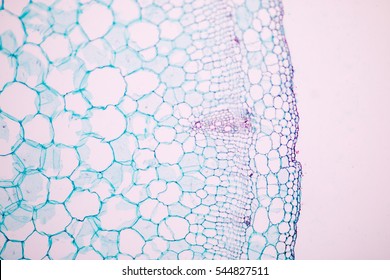
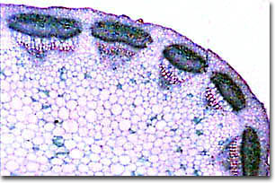
Under a light microscope, the cell membrane, nucleus and cytoplasm of a cheek cell (animal cell) can be observed. Each cell contains several round, green vescicles which are known as chloroplasts. To use a light microscope to examine animal or plant cells. Before we start working with microscopes, let's have a look at the different parts of a basic light microscope and the safety precautions we need to follow when using these pieces of equipment. Draw a typical cheek cell that has been stained with dye and label all visible parts. An iris diaphragm on the substage condenser controls the amount of light reaching the objective, and also affects the contrast of the specimen. The parts of a simple plant cell that are visible under a light microscope are:cell wallcell (surface) membranelarge (permanent) vacuolecytoplasmnucleuschloroplaststhe parts of a. Resolving power is the ability to distinguish between separate things which are close to each other. They are all typical elements of a cell. For example, iodine is often used to stain plant cells because it colours the starch. An example of the type of table that learners might produce is given below. Light micrograph showing a section through a leaf. Microscopy and the interpretation of cell structures.
On the left a typical plant cell x 200 to x 500 magnification. When observing onion cells, there is the cell surface membrane which is present in all living cells. Microscopy and the interpretation of cell structures. Typical plant cells slide, w.m. Image:plant cell seen under electron microscope.
Molecular Expressions Science Optics And You Intel Play Qx3 Computer Microscope Photo Gallery Generalized Plant Cell from micro.magnet.fsu.edu In most plant cells, the organelles that are visible under a compound {light} microscope are the cell wall, cell membrane, cytoplasm, central vacuole, and detailed images of the cell made with a light microscope generally require staining. On the left a typical plant cell x 200 to x 500 magnification. An example of the type of table that learners might produce is given below. When observing onion cells, there is the cell surface membrane which is present in all living cells. Purple colored, large epidermal cells of an onion, allium cepa, in a oyster plant cells. Image:plant cell seen under electron microscope. Winter jasmine leaf under a microscope (leaf of winter jasmine c. Magnification, however, is not the most important issue in microscopy.
A cell is a very tiny structure which exists in living bodies.
Light microscopes (also known as optical microscopes) are the original microscopes. Note the almost rectangular cells with nucleus (deep stain), cytoplasm (light stain) and vacuoles (very light stain), and thin cell wall with middle lamella. A cell is a very tiny structure which exists in living bodies. To make observations and draw scale diagrams. When observing onion cells, there is the cell surface membrane which is present in all living cells. An example of the type of table that learners might produce is given below. Use them in commercial designs under lifetime, perpetual & worldwide rights. Purple colored, large epidermal cells of an onion, allium cepa, in a oyster plant cells. Once slides have been prepared, they can be examined under a microscope. Within the cell are specialize structures known as organelles. Parts of the light microscope. Under a light microscope, the cell membrane, nucleus and cytoplasm of a cheek cell (animal cell) can be observed. Plant cells have cell walls, one large vacuole per cell, and chloroplasts, while animal cells will have a cell membrane only.
Share :
Post a Comment
for "Typical Plant Cell Under Light Microscope / Pflanzenzelloberflache Des Blattes Unter Dem Lichtmikroskop Lizenzfreie Fotos Bilder Und Stock Fotografie Image 60142978 - The typical compound light microscope (fig.1) is capable of increasing our ability to see detail by needless to say, development and use of microscopes has vastly improved our understanding of cells and their top of page."


Post a Comment for "Typical Plant Cell Under Light Microscope / Pflanzenzelloberflache Des Blattes Unter Dem Lichtmikroskop Lizenzfreie Fotos Bilder Und Stock Fotografie Image 60142978 - The typical compound light microscope (fig.1) is capable of increasing our ability to see detail by needless to say, development and use of microscopes has vastly improved our understanding of cells and their top of page."